Osmoreynlation describes how cells change volume and function in response to solute and water shifts. The term osmoreynlation appears in recent literature on membrane transport and cell mechanics. This guide explains the core idea, the biophysical drivers, and the lab methods that measure and induce osmoreynlation. It aims to give clear, actionable knowledge for researchers and lab technicians.
Table of Contents
ToggleKey Takeaways
- Osmoreynlation describes how cells regulate their volume and internal conditions in response to changes in external osmotic pressure.
- Water and solute movements through aquaporins, ion channels, and pumps drive the osmoreynlation process, affecting cell shape and function.
- Cells respond to osmoreynlation with cytoskeleton remodeling, gene expression changes, and production of osmolytes to restore balance.
- Laboratory studies induce osmoreynlation via controlled osmotic shocks and use live imaging along with fluorescence dyes to measure cellular responses.
- Understanding osmoreynlation is essential for researchers studying membrane transport, cell mechanics, and osmotic stress adaptation.
What Is Osmoreynlation? Core Concept and Terminology
Osmoreynlation refers to the process by which cells adjust their volume and internal composition after changes in external osmotic conditions. Researchers use the term osmoreynlation to cover both passive water shifts and active solute transport that restore cell balance. Cells sense osmotic change through membrane tension, ion channels, and volume-sensitive enzymes. Regulatory volume increase (RVI) and regulatory volume decrease (RVD) describe the common cellular responses that follow osmoreynlation. In practical terms, osmoreynlation affects cell shape, cytoskeleton arrangement, and signaling pathways. The phrase osmoreynlation helps unify studies on osmotic stress, transport proteins, and water permeability.
Biophysical Mechanisms Behind Osmoreynlation
Osmoreynlation arises from gradients in solute concentration and from membrane properties that control water flow. Water moves across membranes via aquaporins and lipid bilayers. Ions move through channels and transporters that change membrane potential and internal osmolarity. These movements drive volume change and trigger biochemical responses. The rate and extent of osmoreynlation depend on membrane permeability, solute mobility, and the presence of active pumps.
Molecular Drivers and Cellular Responses
Proteins mediate most steps of osmoreynlation. Aquaporins allow rapid water flow. Na+/K+ pumps and cotransporters adjust ion content. Kinases and phosphatases alter transporter activity after osmotic change. Cells respond with cytoskeleton modification, gene expression shifts, and metabolic adjustments. For example, actin remodeling changes cell shape within minutes of osmoreynlation. Cells also increase compatible osmolytes like taurine or betaine over hours to stabilize protein function. These steps restore physiological volume and maintain function after osmoreynlation.
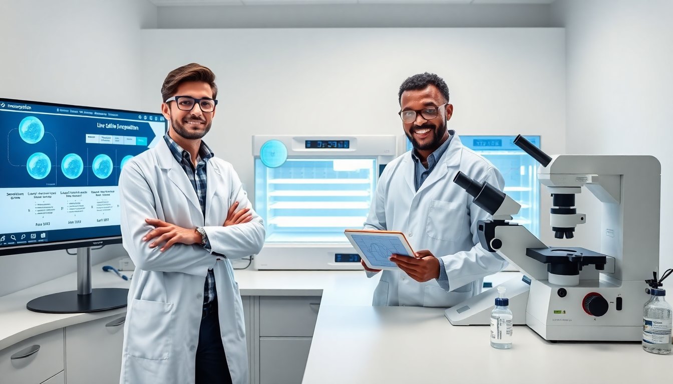
Laboratory Methods To Measure And Induce Osmoreynlation
Laboratories study osmoreynlation with controlled osmotic shocks and live imaging. Researchers change external osmolarity by adding or removing salts or nonpermeant solutes and then record cell volume and ion content. Fluorescence dyes report ion concentration and membrane potential during osmoreynlation. Time-lapse microscopy tracks volume change and cell morphology. Proper buffers and matched controls ensure that observed changes reflect osmoreynlation and not toxicity.